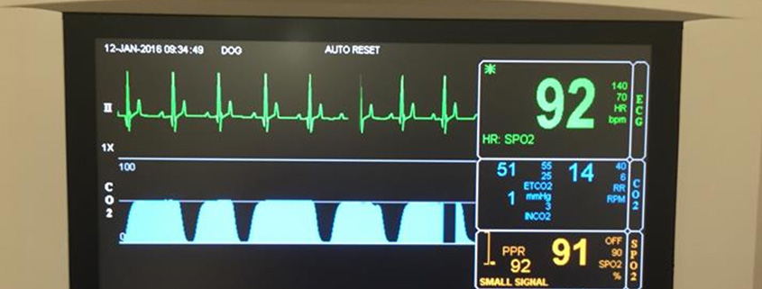
normal end tidal co2 dog
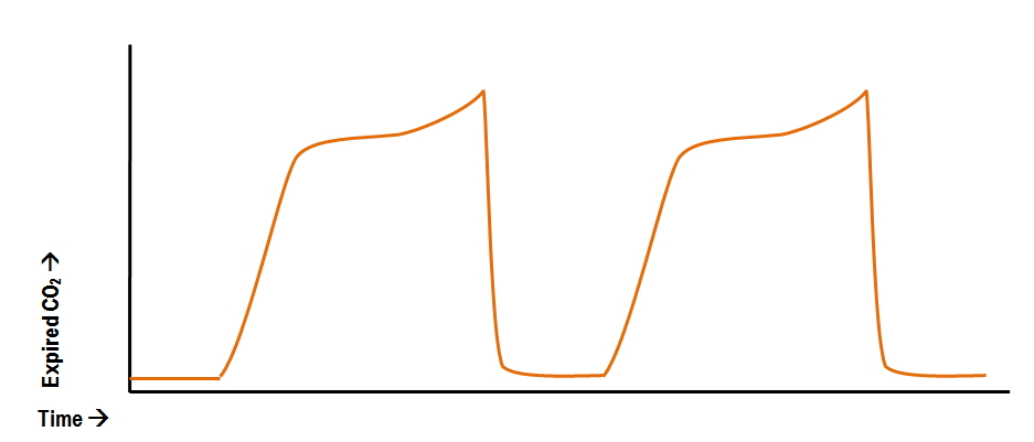
Carbon dioxide during ventilation. A more complete picture of carbon dioxide transfer can be obtained from a capnogram similar to an ECG tracing.

This Article Discusses Why We Should Be Monitoring End Tidal Co2 And How To Troubleshoot Abnormal Values Vettech Veter Vet Medicine Vet Tech School Vet Tech
48 When a person is breathing out CO 2 the graph goes up.
. Capnography waveforms etCO2 and breathing patterns. The arterial to end-tidal PCO2 difference PaCO2-PECO2 was measured in five anaesthetized dogs during controlled ventilation at 025 Hz 15 bpm and during high frequency jet ventilation at 1 3 and 5 Hz. The end-tidal level of carbon dioxide is generally less but is reflective of carbon dioxide in arterial blood and can serve as an indirect noninvasive method of assessing the adequacy ventilation.
What Is A Normal End-tidal Co2 Dog. The capnometer is checked and is in good working order. There was an excellent linear correlation b.
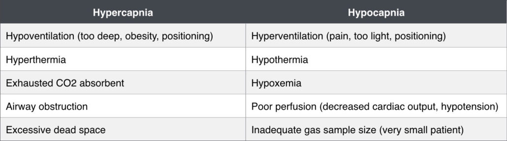
Since problems with lungs are not common and gas exchange between alveoli and the blood is swift and effective. 2 See Figure 1 p. Eased etCO2 levels in cats and dogs are 32 to 35 mm Hg and 35 to 46 mm Hg respectively.
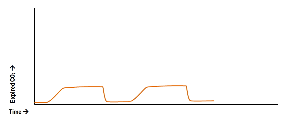
More normal trace with P E CO 2 levels at or above normal. Which of the following is the normal ETCO2 end-tidal carbon dioxide level in an anesthetized dog or cat. Because of the slow response of the infra-red carbon dioxide analyser satisfactory recordings of end-tidal carbon dioxide could not be obtained at.
End-tidal CO 2 monitoring is a non-invasive means of estimating arterial CO 2. The end-tidal carbon dioxide tension PetCO2 measured after a single large tidal-volume breath 15 mlkg body weight was compared to simultaneous measurements of PaCO2 in 6 dogs with normal lungs who were receiving high-frequency jet ventilation HFJV. For example a patient in DKA may have a very low EtCO2.
Well the EtCO2 value is simply a number. 2 to near normal normal EtCO 2 35-45 mmHg represents marked increase of CO 2 delivery to lungs suggesting ROSC If patient develops an organized rhythm after VFVTasystole check EtCO 2 to see if ROSC has occurred CONFIRM PLACEMENT OF ETT After intubation if ETCO 2 10mm Hg tube in trachea. However any number of conditions can cause a change that may or may not be considered normal for any given patient.
Normal range is 35-45mmHg and roughly correlates with the partial pressure of CO2 in arterial blood remember that PaCO2 is usually slightly higher than ETCO2 by 2-5mmHg. Capnography also measures and displays the respiratory rate. Normal minute ventilation about 200 mlkgmin for dogs and cats in conscious animals with normal lungs results in an arterial and therefore alveolar co 2 partial.
In conditions of normal breathing 6 Lmin 12 breathsmin 500 ml for tidal volume etCO 2 is very close to alveolar CO2. During surgery an anesthetized 4-year-old mixed-breed dog has an expired CO2 level of 25 mm Hg. The normal levels of expired CO2 in dogs and cats should between 35 and 45 mm Hg millimeters of mercury.
End-tidal carbon dioxide ETco 2 monitoring provides valuable information about CO 2 production and clearance ventilation. The capnograph is the waveform that shows how much CO 2 is present at each phase of the respiratory cycle and it normally has a rectangular shape Figure 1. End tidal co 2 monitoring is represented as a number and a graph on a monitor.
Yes the generic normal is considered 35-45. Simultaneous comparison of heart rate ECG or stethoscope with pulse rate palpation or blood pressure monitor allows the anesthetist to pick up some dysrhythmias. 4 to 5 CO2 PetCO2 vs.
Under most circumstances healthy pet no chest surgery end-tidal CO 2 is typically 5 10 mmHg less than arterial CO 2. PaCO2 PetCO2 End tidal measurement from expired or exhaled air PaCO2 Arterial blood gas sample End tidal normally 2-5 mmHg lower than arterial Comparing Arterial and End-tidal CO2 Review of Airway Confirmation Visualization Auscultation. Levels that deviate from this range require quick evaluation to determine the appropriate corrective course of action.
Throughout the breath cycle. Also called capnometry or capnography this noninvasive technique provides a breath-by-breath analysis and a continuous recording of ventilatory status. The plateau observed at the end of the.
The CO 2 waveform is a valuable tool for detecting leaks in the anesthetic system rebreathing of CO 2. The measurement of end-tidal carbon dioxide ETCO 2 allows the continuous monitoring of the adequacy of ventilation and circulation in the anaesthetised patientIt measures inspired and expired carbon dioxide CO 2 throughout the whole respiratory cycle using infrared spectroscopyETCO 2 can be of value in the assessment of ventilation metabolism and of a. Heart rate can be monitored from an ECG from a stethoscope esophageal stethoscope pulse oximeter or blood pressure monitor.
Negative Epigastric sounds Equal lung sounds Esophageal detector. In fact its commonly called the ventilation vital sign. Repiratory rate and depth tidal volume which determine minute ventilation and therefore arterial co2.
When a person is breathing in it. Capnography can be used to measure end-tidal CO 2. In normal conditions CO2 is 5 to 6 which is equivalent to 35-45 mmHg.
Repiratory rate AND depth tidal volume which determine minute ventilation and therefore arterial CO2. Which of the following is most likely to correct the hypocarbia. When etCO2 is greater than 40 mm Hg hypercapnia is present more than normal CO2.
If P E CO 2 levels are not normal cardiac output may still be low. The normal end-tidal capnography wave form is basically a rounded rectangle. Monitoring of end-tidal carbon dioxide EtCO2 is a noninvasive method that measures the partial pressure or maximal concentration of carbon dioxide CO2 at the end of exhaled breath which is expressed as a percentage of CO2.
Normal minute ventilation about 200 mlkgmin for dogs and cats in conscious animals with normal lungs results in an arterial and therefore alveolar CO 2 partial pressure of 35 to 45 mm Hg. We know that elevated ETCO2 hypercapnia occurs during hypoventilation and a decrease in ETCO2 hypocapnia occurs with hyperventilation. This is end-tidal CO 2 ETCO 2 which is normally 35 to 45 mm Hg in dogs and 28 to 32 mm Hg in cats.
As spontaneous cardiac function returns CO2 is then delivered to the lungs again and the capnogram will return to more normal levels sometimes P E CO 2 may be high than normal.

Learn More With This Respiratory Article By Melissa Marshall

Learn More With This Respiratory Article By Melissa Marshall

Capnography Bsava2012 Vin

End Tidal Carbon Dioxide Tension Pet Co 2 In Normal Individuals Over Download Table

Did You Know Hypercapnia Is Synonymous With Hypoventilation Pet Health Care Pet Health Veterinary Care
2

Learn More With This Respiratory Article By Melissa Marshall

Learn More With This Respiratory Article By Melissa Marshall
2

Pin On Veterinary
Abnormal Capnography Waveforms And Their Interpretation Deranged Physiology

E Learning Basics Of Capnography Youtube
2

Veterinary Specialist Services Veterinary Anesthesia

Learn More With This Respiratory Article By Melissa Marshall
Riding The Wave Of Capnography Understanding Etco2 Vetbloom Blog
Abnormal Capnography Waveforms And Their Interpretation Deranged Physiology

Learn More With This Respiratory Article By Melissa Marshall
Riding The Wave Of Capnography Understanding Etco2 Vetbloom Blog